Loading video…
All breakouts
10.4Talking HeadHow-To hookHealth
Hook
I want to separate short here or short here
@disasterhbb
163.7K followers · usually 21.8K views a post
Views
2.4M
Likes
39.0K
Comments
476
Shares
2.1K
Why it worked
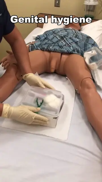
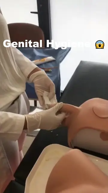
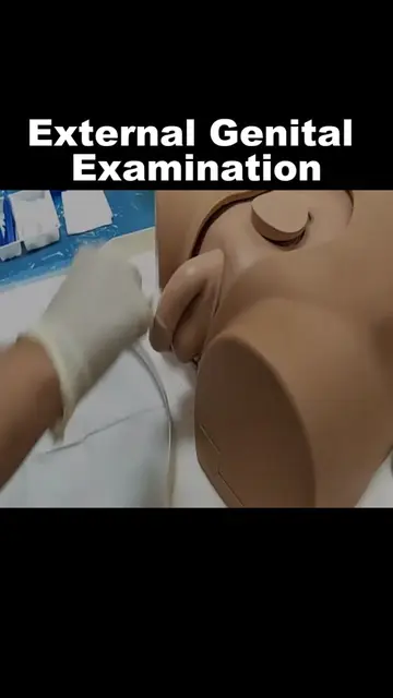
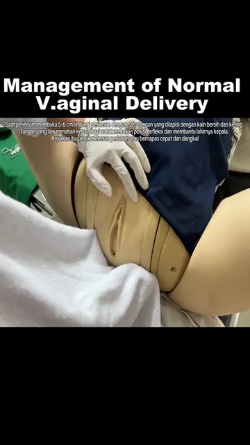
The video uses a clear, step-by-step demonstration on a medical mannequin, making a complex procedure understandable. The use of on-screen text and clear verbal explanations caters to a learning audience, while the close-up shots and focus on tactile feedback provide a sense of realism and engagement.
Summary
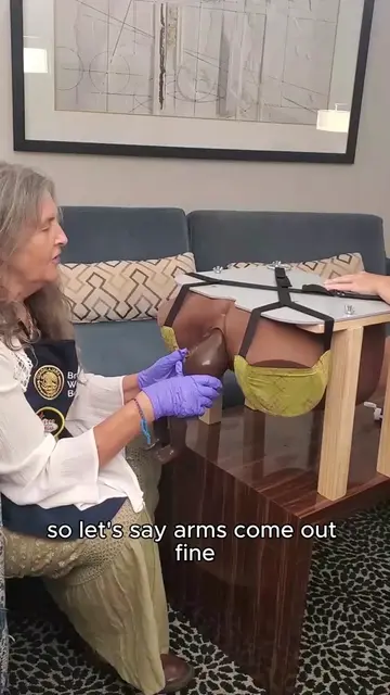
A medical educator demonstrates a procedure on a mannequin, explaining the steps involved in separating tissue and identifying anatomical landmarks. The demonstration focuses on tactile feedback and precise movements to locate the "head" of the fetus during a simulated birth.
Structure
- 1Introduction to the procedure
- 2Identifying separation points
- 3Determining when to cut
- 4Locating anatomical structures
- 5Simulating fetal head presentation
- 6Tactile feedback and finger placement
On-screen text
I want to separate short here or short here
but when do I have to cut
when in the room
flat when it is already in an exclusive period
close-up second shot
third shot left
fourth plane that is already perineating
that I did perineve
as we are going to see,
it is already opening
and it is already like
this
okay, it's already there
turn off your cell phone
for a while
and there goes that's
the little head ah ya
eso
you are touching your
bladder no I don't want
your bladder
I want the head yes
doctor and that's it
from the bud to the head
that
Original caption
#medicaleducation #healthcare #LearnOnTikTok #fyp #medschool
More from @disasterhbb
More breakout videos from this creator.