Loading video…
All breakouts
488Text-HeavyContrarian hookHealth
Hook
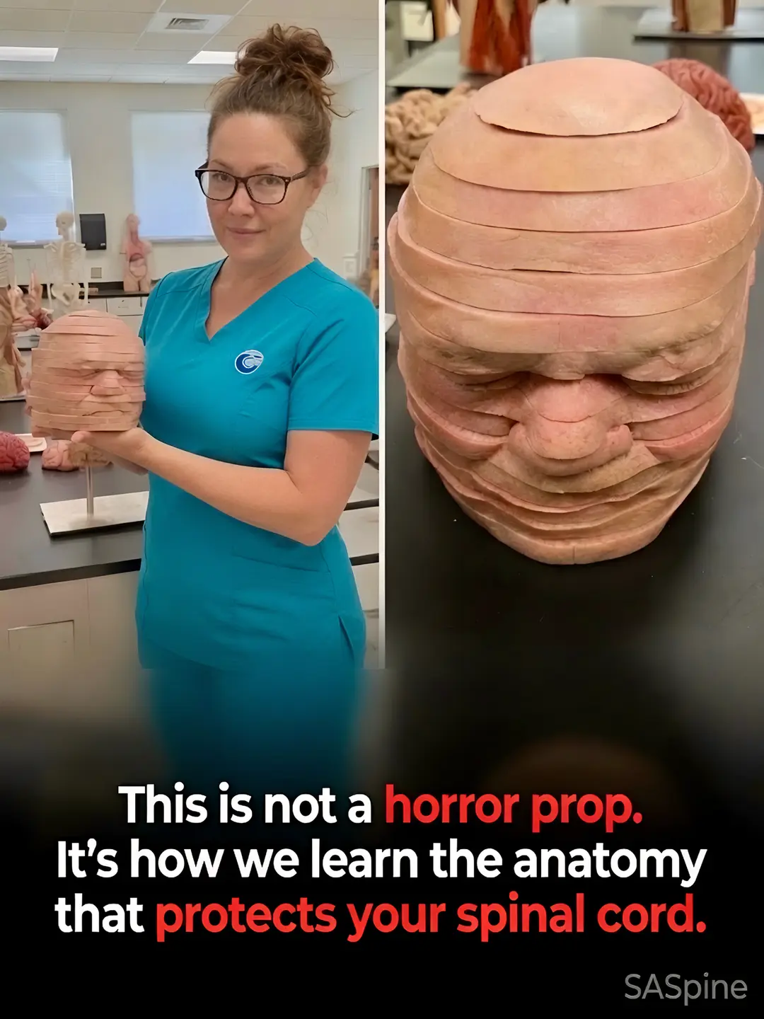
This is not a horror prop.
@stevencyrmd
166.2K followers · usually 1.3K views a post
Views
720.8K
Likes
18.4K
Comments
191
Shares
666
Why it worked
The post uses a visually striking and slightly unsettling image to grab attention, then immediately reframes it with an educational and informative explanation, creating curiosity and providing value.
Summary
The post explains that a sliced head model is not a horror prop but an anatomical teaching tool used to learn the anatomy that protects the spinal cord. It highlights how medical professionals use these models to understand the relationships between different structures in the head and neck.
Structure
- 1Introduction of the anatomical model
- 2Explanation of its purpose in learning spinal cord anatomy
- 3Description of how the model is used for detailed study
- 4Comparison to CT and MRI scans
Product placement
SASpine: It APPEARS as a watermark/logo at the bottom right of the image. Removing it would not change what happens in the video.
On-screen text
This is not a horror prop.
It's how we learn the anatomy
that protects your spinal cord.
SASpine
Original caption
Most people only see the outside of the head and neck. Spine surgeons study it like this. This is an anatomical teaching model used in medical education — sliced into horizontal sections so students and surgeons can understand the exact relationships between bone, disc, nerves, vessels, and soft tissue. Students and surgeons can pull the individual slices apart and reassemble them to study the exact relationships between structures — the same cross-sectional view that appears on CT and MRI scans. Here’s what that level of anatomical detail means for your cervical spine: 1. The spinal cord and nerve roots sit in a very tight space. Small changes in disc height or alignment can create big symptoms. 2. Muscles, ligaments, and fascia work together as a system — treating only one layer often fails. 3. Imaging (MRI/CT) is essentially a high-tech version of these slices. Knowing how to “read” them in 3D is critical for accurate diagnosis. 4. Surgical precision depends on respecting every structure in the field — nerves, vessels, and motion segments. 5. Prevention starts with understanding how posture, load, and movement affect these layered structures every day. At SASpine we use this depth of anatomical knowledge every time we evaluate neck and arm pain, plan procedures, or design non-surgical care. “As a spine surgeon, I’ve spent years studying the neck in this level of detail. These layers are why precision matters so much when patients come in with neck or arm pain.” — Dr. Steven Cyr Have you dealt with neck pain, arm pain, or numbness that didn’t make sense until someone explained the anatomy? Share your experience below — it helps others feel less alone. #CervicalSpine #NeckPain #SpineHealth #SpineSurgery #Anatomy
More from @stevencyrmd
More breakout videos from this creator.