Loading video…
All breakouts
153Text-HeavyDirect Question hookHealth
Hook
WHY TWO X-RAY VIEWS MATTER
@stevencyrmd
165.7K followers · usually 2.1K views a post
Views
178.0K
Likes
5.0K
Comments
20
Shares
74
Why it worked
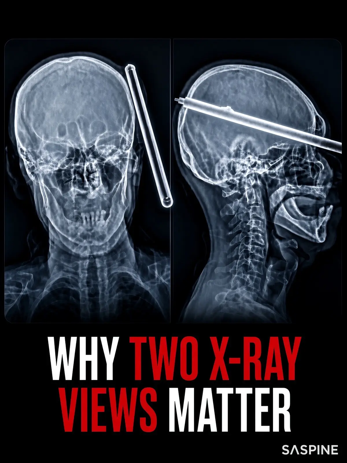
The use of a dramatic and easily understandable visual example (the skull X-ray) effectively illustrates the core message about the importance of multiple perspectives in medical imaging.
Summary
This slideshow explains the importance of using multiple X-ray views in medical imaging, particularly in spine surgery. It uses an example of a skull X-ray to illustrate how a single view can be misleading, while two views can reveal the true situation.
Structure
- 1Introduction to the problem of misleading X-rays
- 2Example of a skull X-ray with two views
- 3Explanation of the risk in spine surgery
- 4Emphasis on the necessity of multiple views
Product placement
SASpine: APPEARS - a logo at the bottom right of the final slide. Removing it would not change what happens in the video.
Call to action
SASpine.com
On-screen text
WHY TWO X-RAY VIEWS MATTER
SASPINΕ
Original caption
One incomplete X-ray can conceal a critical injury. At first glance, the image on the right looks catastrophic—a long metal object appears to have pierced straight through the patient’s skull. But the frontal AP view reveals the truth: the object was lying beside the head the entire time and never entered the brain. In spine surgery, the same risk exists every day. A single view can conceal hidden fractures, subtle misalignments, or instability that only becomes clear from another angle. Relying on incomplete imaging can lead to the wrong diagnosis—and the wrong treatment. That’s why radiographic examinations require at least two different views. An X-ray is only two-dimensional. Overlapping structures create dangerous illusions when seen from only one projection. One view suggested catastrophic trauma. The second revealed reality. “In spine surgery, the first image is never the whole story. We look from every angle before we act.” — Dr. Steven Cyr, Mayo Clinic-trained dual-trained orthopaedic and neurosurgical spine surgeon At SASpine, we never settle for incomplete information. If you’re dealing with neck or back pain, unexplained symptoms, or were told “everything looks fine” on a single X-ray, schedule a comprehensive evaluation with our team. Accurate multi-view imaging is the first step to the right diagnosis and the right care. #SpineSurgery #SpineHealth #Radiology #NeckPain #BackPain
More from @stevencyrmd
More breakout videos from this creator.